
Diseases
Retinal detachment
Definition
Retinal detachment is a serious condition which, if left untreated, can lead to loss of sight.
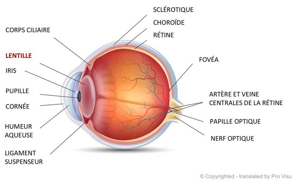
The retina is the membrane that lines the back of the eye. It receives images and transforms the beam of light received into an electrical signal so that it can be sent to the brain. As we age, the vitreous body (the gelatinous substance that fills the inside of the eye and is in contact with the retina) liquefies, contracts and can pass behind the retina, no longer allowing it to adhere to the back of the eye. This tensile force can then suddenly tear or even detach the retina.
Causes
There are several risk factors:
- Age, because as we age, the retina thins. The risk of retinal detachment increases from the age of 40 and is considered significant from the age of 50.
- Myopia, because the retina of people with myopia is tighter, which increases the risk of tears.
diabetes - Eye injuries
- Cataract surgery
- Family history
Symptoms
Retinal detachment is painless, but there are symptoms that can precede its appearance. The various vision problems that may indicate retinal detachment are as follows:
- Shadows, black or grey, called floating bodies: sometimes referred to as flies or filaments, which suddenly appear in the field of vision.
- Luminous images or flashes, known as phosphenes: sufferers describe flashes when they move their eyes
- A more or less dark curtain that masks part or all of the field of vision (this discomfort increases as the detachment progresses)
- A reduction in vision
- A dark veil at the periphery of the visual field, which will widen towards the centre
As soon as the macula (the centre of the retina) is affected, the detachment causes a significant and rapid decline in vision.
Diagnosis
The diagnosis of a detachment requires a precise examination of the retina: the fundus. To ensure that the examination is carried out correctly, the pupil is dilated. The doctor can then examine the retina using a slit lamp. In some cases, an ultrasound scan may be necessary to get a better look at the back of the eye.
The ophthalmologist also checks visual acuity and field, as well as the pressure in the eye.
Treatment
Retinal detachment is generally synonymous with a medical emergency. It requires surgery within a short space of time, from a few hours to a few days. However, if the detachment begins and only tears are present, without any real detachment, laser treatment may be sufficient.
Surgery removes the liquid responsible for the detachment, which has infiltrated between the retina and the back of the eye, and then repairs the tears. There are various techniques, depending on the severity and type of detachment, but the most commonly used is vitrectomy. This involves removing the vitreous body and sealing the tear with a laser.
The retina is then held against the wall of the eye using gas or silicone oil. The operation may cause some pain in the eye, a feeling of a foreign body and swelling of the eyelids. These symptoms generally subside within a few days.
The effects of an operation vary according to the extent of the retinal detachment, its location and the age of the sufferer. The operation does not always restore normal vision, but rather stabilises it so that it does not worsen. In rare cases, several operations may be necessary.
Frequency
Retinal detachment is a relatively rare disease, affecting one person in 10,000. It mainly affects people over the age of 50 and those suffering from severe myopia or diabetes.
Prevention
There is only one option for trying to prevent the onset of retinal detachment: systematic and regular retinal examinations for people at risk.
As retinal detachment progresses from the periphery of the eye towards the centre, prompt medical attention when the first symptoms appear will enable retinal detachment to be treated quickly and effectively.
References
Le décollement de rétine - Service d'ophtalmologie | HUG - Hôpitaux Universitaires de Genève
decollement_retine.pdf (hug.ch)
Contenu revu et contrôlé le 27.04.2023.